The problem
The cornea is the eye's transparent outer layer that lets in light. It consists of three cellular layers: epithelium, stroma, and endothelium. When you age, your corneal endothelium deteriorates. It slowly fades while endothelial cell density decreases, causing corneal opacity or other corneal diseases. An estimated 12.7 million people with corneal opacity are waiting for treatment worldwide.
Today's state-of-the-art therapy for corneal opacity is transplantation. Although effective, a lack of donors hampers results—there's only one donor available for every seventy patients. Regenerative medicine, the biomedicine area that develops therapies helping the body regenerate tissues such as the cornea, could help meet that demand.

Vanessa LaPointe is an Associate Professor at the MERLN Institute for Technology-Inspired Regenerative Medicine and group leader of the institute's Cell Therapy group. One of their projects is building cell therapies for cornea disease.
"A real challenge in regenerative medicine is defining success," she says.
LaPointe: "In the lab, we're trying to build cell therapies or tissue-engineered therapies to replace diseased or damaged tissue. But for some tissues, like the corneal endothelium, the healthy tissue is not yet completely defined, and the assays associated with it aren't necessarily great. So how do we know if we have engineered a good endothelium?"
She delineates the value of single-cell RNA sequencing in regenerative medicine. "What you need is a benchmark to ensure that what you've engineered in the lab is actually the healthy tissue the patient needs." She explains: "For that, you need a fundamental understanding of what the corneal endothelium is. And the best way to do that is with techniques like single-cell RNA sequencing."
The solution: 10x Genomics Single Cell Gene Expression
LaPointe explains how her team decided to work with Single Cell Discoveries: "First of all, single-cell RNA sequencing is a leading method. Second, it's very fitting for the questions we had: the discovery of new cell type markers, understanding tissue heterogeneity, and benchmarking against characteristics of healthy tissue."
“I found it important as a supervisor that Pere understood what was happening and that he was involved in the process. So it was ideal to have such good people to work with.” – Vanessa LaPointe, group leader
The team considered several other techniques before landing on 10x Genomics Single Cell Gene Expression. "Our idea was to start with SORT-seq," says Pere Català, PhD candidate at LaPointe's Cell Therapy group and first author of the Scientific Reports paper. But in early discussions with Single Cell Discoveries, they came to a different conclusion.
"10x Genomics would make more sense due to the higher amount of cells that could efficiently be sequenced," Català clarifies. "It would also make it easier to prepare your cells. You don't need to FACS-sort cells and put them in wells, just dissociate the tissue and send them fixed in methanol."
LaPointe remembers early discussions with Single Cell Discoveries' team members Nathalie, Judith, and Mauro. "They advised us, 'Maybe, you guys would find it a bit more convenient to do the 10x Genomics protocol.' And they were right."

The challenge
Still, the 10x Genomics protocol is not without trials. Català: "The most challenging part was the high amount of cells you have to get from a single cornea to perform a successful 10x Genomics experiment."
Although Single Cell Discoveries has experience in lowering the number of cells required for 10x Genomics, you still need a couple of hundred thousand cells as input to get high-quality single-cell data on several thousand cells.
"We got asked to provide at least 200,000 cells," Català explains. "For corneal epithelium and stroma, we didn't have any type of problem. But especially for the endothelium, it was a big headache." The corneal endothelium is a relatively thin layer and constitutes small numbers of cells per sample. "So we were trying to work with endothelial suspensions of about 100,000 cells." That was no easy achievement. "We had to work with 10–15 corneas to optimize the dissociation protocols."
After optimization, they could get just enough viable cells for four samples.
The Single Cell Discoveries data analysis team delivered the first experimental results. But not only that—they also found a workaround for the relatively low number of endothelial cells.
The solution: data analysis services
Why did LaPointe and Català use Single Cell Discoveries' data analysis service? LaPointe: "I think we were very, kind of, self-aware that single-cell data analysis was not our expertise. Still, we didn't want to outsource data analysis to the point where you just send a list of tasks to someone and get the answers sent back. I found it important as a supervisor that Pere understood what was happening and that he was involved in the process. So it was ideal to have such good people to work with."
“This work formed the basis for future experiments. We can use the cornea atlas we made and the markers we found to understand how these cells might undesirably change over prolonged culturing periods.” – Vanessa LaPointe, group leader.
"For me," Català adds, "the collaboration with the data analysis service was fantastic. Nathalie had the expertise in terms of data analysis, and I have more biological expertise. So whenever I asked a question, she would propose a solution. Or then, when she was asking a question, I would propose some solution. It was excellent work in tandem."
LaPointe: "I could just send Pere off to these discussion meetings because I could trust the process. And I didn't have to attend myself all the time, which is also wonderful for a PI. Pere could discuss his research aims and get advice from the company on which directions to take and what could lead to something."
Collaboration ultimately solved the team's issues with the low-quality, low–cell count endothelium samples that had become this study's most challenging bottleneck. Català recounts the process: "So, the first separation we did was manually dissociating the cells for each corneal cellular layer. We sequenced each layer separately. Then, when clustering the single-cell data, we knew that each cell came from that specific layer." That gave the clustering for the first four samples high accuracy.
He goes on, "Once Nathalie did the first data analyses, we would see that the cells clustered per layer successfully. That could mean that if we pool the cells from all the layers and sequence them together, we could identify from the data what cells were from what layer."
Pooling the layers for sequencing would considerably increase the cell number, and the chances of a successful 10x Genomics run.
"We decided to sequence two additional samples where we would dissociate the layers separately but pool all the cells for sequencing," Català says. That idea worked.
Català: "We had to ensure that the layer clustering was biologically sound first. But then, the technical decision to pool all cells for sequencing solved our issues."
The results were coming in.

The results
"We were surprised to see so many different cell types," Català begins. "Of course, we were expecting heterogeneity. But identifying nine cell clusters in the cornea epithelium was, for me, kind of striking. Also for the stroma, we identified four cell populations and the identification of a small fibrotic cell population in there was very interesting."
The team also identified two cell clusters in the hard-to-attain data of the corneal endothelium. Català: "Even though it was super challenging to dissociate the cells, we could still identify two cell populations that were rather different. These main findings helped us understand the heterogeneity of the healthy human cornea."
"And besides all that," he continues, "we could identify novel cell markers to locate some subsets of epithelial cells with stem-cell characteristics." The association of these cells in existing treatments for limbal stem cell deficiency gives this finding clinical and technological relevance.
“What we appreciated most was the nature of the collaboration. There are other companies you can send samples to for sequencing. You can send them overseas and get your data sent back. But here, we could co-design the experiments together.” – Vanessa LaPointe, group leader
LaPointe agrees: "This work formed the basis for future experiments. In a study we're doing now, for example, we are isolating corneal endothelial cells from a donor and culturing them to expand them into large numbers for therapy. We can use the cornea atlas we made and the markers we found to understand how these cells might undesirably change over prolonged culturing periods. This is relevant to regenerative medicine's aim to develop cell therapies."
"This kind of discovery that helps to understand what happens in healthy tissue," LaPointe concludes, "is really important for defining a success in regenerative medicine."
Future projects
The Cell Therapy group and Single Cell Discoveries are working together again on some of these current studies.
In today's research climate, that's a sensible decision, LaPointe explains: "There's so much emphasis now on public-private partnerships in the grant application process. So it's a very logical thing to bring companies that we're really happy working with into the consortia we're building."
"What we appreciate most is the nature of the collaboration," she elaborates. "There are other companies you can send samples to for sequencing. You can send them overseas and get your data sent back. But here, we could co-design the experiments together. Research is never just a formula. You have many decisions to make. With Single Cell Discoveries, we could discuss the experimental design, which analyses to do and how to solve our challenges."
"Of course, most things never work for the first time," Català adds, "but the solutions ultimately were there."
LaPointe: "That's, I think, really ideal. At the start of a study, we're still figuring out a lot. We don't know exactly what we're looking for when we start these experiments. So you need that kind of collaborative relationship to get the most out of big datasets. And if I look back to the original ideas we had for this work, it's not nearly as good as what it turned out to be. That was really through the discussions with the team of Single Cell Discoveries. It definitely made a better paper."
You can read the paper here.
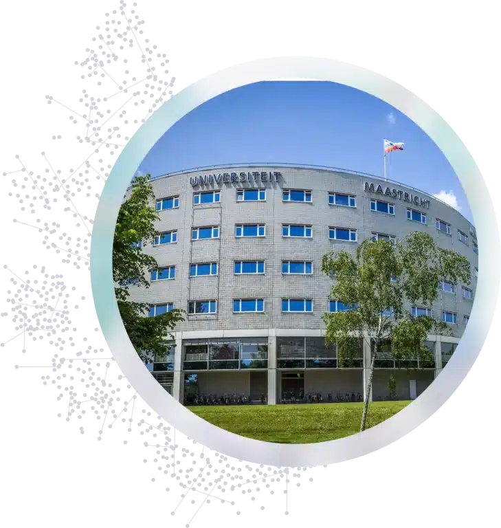
Photograph of the MERLN Institute by Marcel van Hoorn.