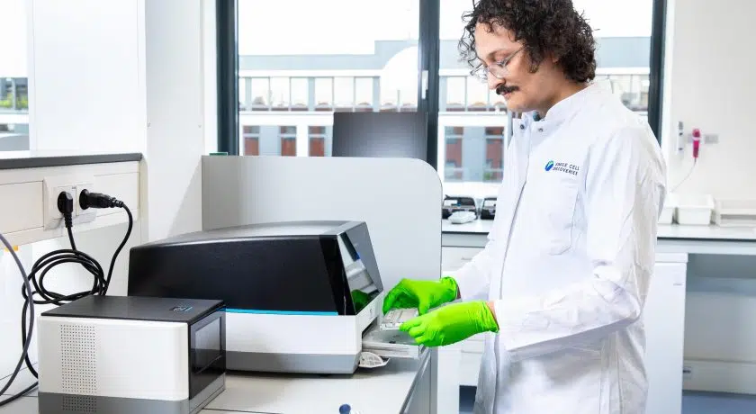
Spatial transcriptomics is an overarching term for all methods that assign transcriptomics data to the original location within the tissue. Here, we explain why to consider spatial transcriptomics and from which spatial techniques you can choose.
A brief history of spatial transcriptomics
Although the term spatial transcriptomics was first introduced in 2016, the first steps were already taken in the late ’60s with the use of in situ hybridization. This is a technique that stains nucleic acids at their original location within the cell or tissue. This was followed in the late ’90s by the first microdissection techniques, in which a microscope is used to dissect a small portion of tissue.
A new wave of techniques started to emerge around 2012 in an effort to combine spatial information with single-cell sequencing. This was with great success.
All this work resulted in spatial transcriptomics being titled by Nature as “Method of the Year 2020”.
Why spatial transcriptomics?
By collecting transcriptomic data, scientists are able to assign “traits” to the studied cell types, as the transcriptome reflects which genes are actively being up- and downregulated. With the development of single-cell RNA sequencing, it became possible to measure transcriptomes at a single-cell resolution. This was a huge achievement of the scientific community. Despite its impact, single-cell RNA sequencing has an important drawback, as tissues are typically dissociated into single cells and the cells are taken away from their context within the tissue.
Why is that important?
Each cell is influenced by its direct surroundings. So, if we understand what is surrounding a particular cell, it helps us understand why a cell is responding in a certain matter. With spatial transcriptomic, it is possible to obtain information on the transcriptomes of a single cell or a small group of cells, while maintaining the information on where the cell (or group of cells) is located within the tissue.
Techniques for spatial transcriptomics
There are roughly four groups of methodologies to conduct spatial transcriptomics. These groups all have a large variety of techniques. It is good to keep in mind that not all spatial techniques have a single-cell resolution, or provide information on the whole transcriptome.
1. Microdissection
Microdissection is a technique where you isolate regions of interest from within a sample. Isolation is done by lasering or cutting. From these tissue pieces, RNA is isolated, processed, and sequenced.
2. Fluorescent in situ hybridization
Fluorescent in situ hybridization is a spatial transcriptomics method to visualize RNA at the original location within the cell. This does not involve sequencing and therefore does not provide information on the full transcriptome. However, these techniques can often achieve a high resolution.
3. In situ sequencing
In situ sequencing is the sequencing of RNA while the cell stays within the tissue context. The sequenced RNAs are visualized using fluorescent probes. This method also does not allow for full transcriptome analysis.
Examples: FISSEQ|Barista-seq
4. In situ capturing
In situ capturing is a spatial transcriptomics method in which transcripts are first captured and barcoded within the tissue. Then, sequencing is performed outside the tissue. The tissue is imaged, which allows the transcriptomics data to be overlayed with the tissue images.
Examples: Nanostring|Visium
5. In silico construction
These methods computationally map single cells back into space, with little or no prior knowledge about the position of the sequenced cells. These methods are therefore compatible with single-cell RNA sequencing.
Example: novoSpaRc
At Single Cell Discoveries, we offer 10x Genomics Visium Spatial Gene Expression as a service. This is an in situ capturing technique to perform spatial transcriptomic. Read more about Visium in our blog “What is Visium?”


