Single-cell spatial transcriptomics pinpoints drug-tolerant niches in colorectal-cancer liver metastasis

A recent photo of PERSIST-SEQ CONSORTIUM members

A new generation of spatial transcriptomics enables researchers to capture gene expression at a 2 × 2 µm resolution, directly linking tissue morphology with whole-transcriptome data. This unprecedented level of detail is transforming how biology is studied in situ, from mapping immune cell organisation to identifying rare, drug-tolerant tumour clones. Visium HD is at the forefront of this shift, redefining what is possible in spatial biology.

Single Cell Discoveries and IRB Barcelona are both members of the PERSIST-SEQ consortium (photographed in the image above). In this case study, we describe our collaboration with the Colorectal Cancer Laboratory at IRB Barcelona. Through our end-to-end spatial workflow service built around Visium HD technology, we supported the identification of distinct micro-niches of colorectal cancer cells that persist after FOLFOX chemotherapy. Together, these insights provide a roadmap for developing more effective combination therapies.

PERSIST-SEQ consortium, IRB & SCD

The goal of the PERSIST-SEQ consortium is to enhance understanding of therapeutic resistance in cancer and to develop effective strategies to improve cancer treatment and prevent drug resistance. As a result of this effort, the project anticipates a significant step forward for the cancer community and its understanding of tumor plasticity.

PERSIST-seq aims to change how scientists and clinicians view cancer and its associated drug development. Within this broader effort, IRB Barcelona leads a work package focused on pre-existing tumor states that persist after treatment.

Team mates from IRB

Adrià Jaume Roura (Left) and Mireia Artola (Right), along with colleagues at IRB Barcelona, study why colorectal cancer metastases relapse after FOLFOX chemotherapy. Using Visium HD on FFPE liver sections, Single Cell Discoveries profiled 6.5 × 6.5 mm areas to better understand cancer heterogeneity during treatment.

High-Resolution Spatial Transcriptomics

Single-cell RNA sequencing reveals extraordinary cellular diversity, but it also disrupts tissue architecture and separates cells from their original locations. Visium HD restores that missing layer: a continuous array of 11 million 2 µm pixels is uniquely barcoded, enabling researchers to overlay micron-scale gene-expression maps directly onto H&E or immunofluorescence images, without having to choose between histology and omics.

Behind the technology: pixels, probes, and poly-A

Visium HD at a glance:

  • Continuous 2 µm resolution: ~25 × finer than first-generation Visium
  • Whole-transcriptome probe sets for human and mouse (18–19 k genes)
  • Automated CytAssist™ glass slide to Visium slide transfer preserves geometric fidelity
  • Compatible with FFPE, fresh- or fixed-frozen tissue
  • NEW 3′ poly-A chemistry for fresh-frozen samples: species-agnostic capture, compatible with spatial TCR/BCR or long-read sequencing

Picture CytAssist as a slide “sandwich”: hybridised probes on mRNA molecules lift off the tissue section, land on the barcoded lawn, and keep their exact x‑y coordinates for sequencing.

Watch our webinar on spatial profiling of drug-tolerant cells in colorectal cancer models with Visium HD

Single-cell spatial insights using Visium HD

WATCH WEBINAR

Why outsource your spatial projects to Single Cell Discoveries?

Spatial assays involve specialised hardware and deep sequencing. SCD makes the leap frictionless:

  • Early-stage design: Power calculations, sample preparation, and sample preservation advice
  • Slide-in, data-out: Ship FFPE or frozen sections; SCD runs the imaging, CytAssist, library prep, and NovaSeq X Plus sequencing
  • Four-to-six-week turnaround: Interactive exploratory report with clusters, differential expression, and spatial overlays
  • Advanced consultancy: Integrate Visium HD with matching single-cell datasets or custom probe panels

“Our goal is to let scientists focus on the biology, not the logistics,” states Oriol Llorà Batlle, Team Lead R&D at SCD. “Because we run the entire pipeline routinely, quality and speed stay high, even for large clinical cohorts.”

Find out how we can help you with your research question

Download a detailed overview of our transcriptomics services

Get our services brochure

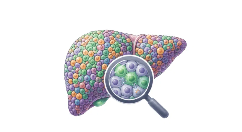
Spatial transcriptomic insights into treatment-resistant colorectal cancer

To capture how chemotherapy reshapes tumour architecture, we performed Visium HD spatial transcriptomics on matched untreated and FOLFOX-treated liver metastases, profiling entire 6.5 × 6.5 mm tissue sections at near-cellular resolution.

Spatial transcriptomics maps of colorectal cancer liver metastases comparing untreated and FOLFOX-treated samples, showing whole-section gene expression coverage across 6.5 × 6.5 mm tissue areas using Visium HD.

Spatial transcriptomic profiling of untreated and FOLFOX-treated colorectal cancer liver metastases. Whole-section Visium HD maps show global transcriptional activity across matched samples, highlighting treatment-induced changes in tumour organisation while preserving spatial context.

Pre-existing survivors
Even before therapy, small epithelial substates expressing LGR5 (a stem cell marker) and EMP1 (a pro-metastatic/EMT marker) can be detected. After FOLFOX, these cells expanded dramatically, indicating that their capacity to tolerate chemotherapy was already encoded in the biology of the untreated tumour rather than newly acquired.

Anatomical hot-spots
Most resistant regions are localised at the tumour rim, right where cancer cells interface with stromal fibroblasts and vasculature, but notably away from dense immune infiltrates. This spatial separation hints at a protective niche that may shelter clones from both drugs and immune attack.

Lower overall diversity
The chemotherapy eliminated many of the initial metastatic sites, resulting in more transcriptionally uniform post-treatment lesions. Yet within that seemingly homogeneous mass, Visium HD still pinpointed isolated resistant pockets: targets that bulk RNA-seq would have missed entirely.

“Visium HD let us trace resistant clones back to their exact position, something bulk or standard Visium could never show,” says Roura. Those coordinates now guide follow-up single-cell and functional assays to test combination therapies that could eradicate the niche.

Why Single Cell Discoveries offers Visium HD as our preferred spatial transcriptomics service:

Key Advantages of Visium HD:

Pathology-grade imaging combined with transcriptomics
High-resolution H&E or immunofluorescence images can be directly overlaid with whole-transcriptome spatial maps on the same tissue section. This eliminates the need to choose between tissue morphology and molecular data, enabling true pathology-grade spatial analysis.

Broad species compatibility
The 3′ poly-A RNA sequencing chemistry captures transcripts from virtually any organism, including human, mouse, and zebrafish. This makes the platform well-suited for developmental biology, comparative genomics, and translational research across species.

Built-in multi-omics expansion
Spatial gene expression can be extended with TCR and BCR profiling or long-read isoform sequencing. These additions allow deeper insight into cell identity, immune clonality, and transcript diversity within a single experiment.

End-to-end service scalability
All CytAssist processing, library preparation, and deep sequencing can be handled by a specialized service team, with results delivered via a secure, scalable analysis infrastructure. This allows rapid turnaround without the need to expand internal laboratory or bioinformatics capacity.

Whether you are deciphering tumour resistance, mapping immune infiltration, or exploring organ development, Visium HD offers a clear window into the where behind the what.

Choosing the Right Method: Technology choices you can trust

Transcriptomics method selection depends on multiple factors, including tissue type, sample preparation feasibility, desired resolution, and budget. SCD supports the design of tailored workflows for Bulk-seq, scRNA-seq, spatial transcriptomics, or integrated approaches.

Our available support includes:

  • Technical expertise across all transcriptomics methods
  • Sample preparation guidance and tissue handling protocols
  • Scalable project design with integrated analysis and reporting
  • Each project is individually evaluated to ensure the chosen method yields meaningful biological insights.

Contact our team to discuss your research goals and explore the best transcriptomics strategy for your study.

Acknowledgement of support
PERSIST-SEQ receives funding from the Innovative Medicines Initiative 2 Joint Undertaking under grant agreement No. 101007937. This Joint Undertaking receives support from the European Union’s Horizon 2020 research and innovation program and EFPIA. See www.imi.europa.eu for more details.

Download our services brochure