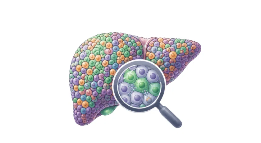
Understanding complex tissues and diseases requires more than a single readout. By combining bulk, single-cell, and spatial transcriptomics with targeted proteomic validation, we can now follow and understand biology from broad patterns to precise cell states and their location in the tissue. This integrated approach enables the identification of meaningful targets, understanding of (tumor) microenvironments, and confirmation of functional mechanisms in a matter of weeks.
This blog discusses how transcriptomics and proteomics complement each other in the single-cell and spatial era, and why integrating these layers is now essential for understanding complex biology.

Jump to a section in this blog:
Mutiomics for complex biology
For years, transcriptomics and proteomics were viewed as separate approaches, each offering a partial view of cell biology. With the rise of single-cell RNA-seq, such as 10x Genomics Flex, Parse Evercode, and many others, and high-resolution spatial transcriptomics platforms like CosMX or Visium HD, that separation no longer makes sense. Modern studies increasingly rely on multi-layered data, RNA and protein, single-cell and spatial, to understand how complex tissues behave and how cells respond to perturbation, and monumental data efforts to tie it all together.
At its core, transcriptomics measures intention: gene expression programs that define cell identity, state, and potential. Proteomics measures action: receptor presence, signaling activity, and post-translational modifications.
Learn more about the full scope of our transcriptomics expertise
Download a detailed overview of our transcriptomics services
RNA: A Cost-Effective, Unbiased View of Cellular Identity
Bulk, single-cell, and spatial transcriptomics remain the most effective approaches for mapping tissue architecture at scale.
- Bulk RNA-seq captures pathway-level differences across large sample sets.
- Single-cell RNA-seq resolves heterogeneity and reveals discrete cell states.
- Spatial transcriptomics places these states back into the tissue with near single-cell resolution.
This transcriptomics layer excels at target discovery, early hypothesis generation, and understanding transcriptional programs underlying development, inflammation, or therapeutic response. RNA is abundant, sensitive, and available across many sample types, including FFPE. Assays to read out RNA levels are, by design, unbiased: they require no a priori knowledge of the biology in question.
But RNA alone cannot establish whether a receptor is expressed at the membrane, whether a checkpoint is active, or whether a pathway is engaged at the protein level.
Protein: The Functional Layer Driving Cellular Mechanisms
Proteomics fills the gaps that transcriptomics can’t address.
Single-cell proteomics (generally mass spectrometry or Antibody-based) and spatial proteomics (Circular Fluorescence or Mass spectrometry-based) quantify receptors, signaling nodes, cytokines, and post-translational modifications. These are the readouts that define functional cell states.
Protein-level information is essential for:
- distinguishing primed vs activated immune states
- assessing signaling pathway activity
- verifying membrane-bound targets
- confirming ligand–receptor interaction
- translating transcriptomic findings into real-world target validation
Proteomics complements RNA by testing whether transcriptional predictions lead to biochemical action. In the end, we usually don’t want to drug an RNA, but a protein.
Why Single-Cell and Spatial Must Be Read Together
Single-cell assays provide resolution but remove tissue structure. Spatial assays preserve architecture but often trade off depth or modality. Used together, they reveal:
- which cells exist and in what states
- where they are located in the tissue
- which microenvironments shape them
- how cell–cell interactions form functional niches
- how drug response changes the local organization of cell types
High-definition spatial platforms, such as 10x Genomics' Visium HD or Nanostring CosMX, increasingly enable researchers to interpret single-cell data with anatomical precision. Meanwhile, single-cell technologies like 10x Genomics or (more recently) Parse Evercode extend single-cell-like profiling to fixed tissues and hard-to-dissociate samples, aligning datasets across modalities. When transcriptomic and proteomic signals agree in both single-cell and spatial contexts, confidence in mechanisms rises sharply, and de-risked targets can be found and validated quickly.
There are, however, some instances that benefit from choosing one of the two modalities. To learn more about when to choose single-cell sequencing, spatial transcriptomics, or both, please read this blog.
Watch our webinar on spatial profiling of drug-tolerant cells in colorectal cancer models with Visium HD
Single-cell spatial insights using Visium HD
A Practical Multi-Omic Workflow for immuno-oncology
With the advent of FFPE-compatible transcriptomics across bulk, single-cell, and spatial, we are often employing the following strategy to deliver good targets quickly:
- We start with Bulk RNA-seq on dozens or hundreds of FFPE samples to screen for pathway shifts or drug response signatures. This allows us to identify interesting candidate samples quickly and at a relatively low cost.
- Single-cell RNA-seq, such as 10x Genomics Flex, on a sample subset, allows us to resolve the cell states driving those signatures. This gives us precise insight into whether the right cell types are responding to, say, the drug of choice, and helps us pick the right samples for follow-up analysis.
- Spatial transcriptomics, such as Visium HD, can then be done on the most informative tissues to see where these states reside and how they organize within the tumor or tissue microenvironment. This layered transcriptomics approach delivers partially derisked targets in a short amount of time.
Single-cell or spatial proteomics can then be used to validate and confirm functional markers such as receptor abundance, checkpoint expression, or immune infiltration. Setting up a proteomics method that is relatively flexible and can handle different proteins/targets to validate each iteration is key: otherwise, target finding outpaces validation. This pipeline is compelling in immuno-oncology, where knowing which immune cells infiltrate the tumor and whether their receptors and signaling pathways are active cannot be answered by RNA alone or by single-cell or spatial data alone.
But while this is a strong example, the general principle holds across many fields: RNA finds the candidates; protein confirms the biology. Spatial ties the story together.
Where the Field Is Going
As resolution improves and workflows mature, biology is moving toward coherence across modalities:
- Transcriptomics for identity and potential
- Spatial assays for context and de-risking the right targets
- Proteomics for function, mechanism, and validation
Integrative models that treat these as complementary rather than competing methods. A big task remains in fully integrating these effectively into a single data pipeline, rather than relying on complex Venn diagrams after separate analyses. AI has been suggested as the leading solution. The responsibility of layering several layers of omics data in the correct order, and filtering through the false positives NGS data produces with well-written code, will remain with humans - for the time being, at least.
The goal is not to choose between transcriptomics and proteomics but to understand how each layer strengthens the other. When a cell state is defined transcriptionally, validated functionally, and located precisely in tissue, that signal becomes robust, fit for discovery, for mechanism, and for translational decision-making.
In this sense, the future of single-cell and spatial biology is not multi-omics by fashion, but multi-omics by necessity. Only by combining layers do we approach the biological truth we’re trying to measure.